digital radiology of exceptional quality
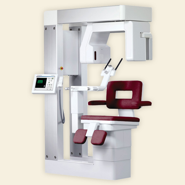
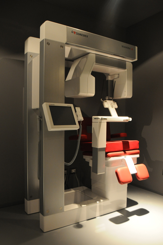
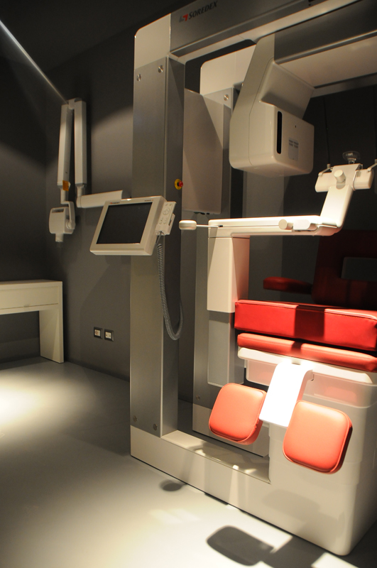
Scanora 3D
Three-dimensional digital multifunctional radiology system for integral images of the dento-maxillo-facial region and dedicated panoramic views.
CBCT Cone Beam*
- Excellent and high precision image quality. Images are acquired in high definition for detailed evaluations or in standard definition for routine work.
- Low radiation dose to the patient.
The 3D images clearly show dental anatomy and bone structure, and enable the visualization of the soft tissues of the entire maxillofacial region.
The operating software is based on a very sophisticated algebraic volumetric reconstruction, ART, which improves image quality, eliminating the problems deriving from the presence of artefacts such as: fillings, implants, bridges, crowns, or the patient’s movements during acquisition.
* CT cone beam has become more and more important in treatment planning and diagnosis.
This technology is increasingly being used in implantology, and in particular in computer assisted implantology techniques, but also in other dentistry fields, such as endodontics and orthodontics.
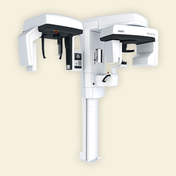
KaVo OP 3D Pro
Digital radiological device equipped with acquisition functionality in panoramic, cephalometric and 3D (CBCT) mode, for images of the dento-maxillofacial region.
It guarantees extreme comfort for both the patient and the operator, high quality diagnostic images and low dose administered to the patient, thanks to the patented Low Dose Technology ™ ". For clinical cases that are particularly sensitive to dose, such as post-operative investigations or pediatric exposures, the reduction of radiation represents a benefit of fundamental importance.
Correct positioning of the patient is guided by reference laser lights that turn on automatically. Stable patient positioning via 5 support points reduces patient movement. The open design allows for a simple and accurate view.
As for three-dimensional images, KaVo OP 3D Pro offers the possibility of acquiring up to five different volumes, each with three image resolutions. Each setting provides the perfect resolution for the specific diagnostic question. The five different volumes guarantee a diagnosis Accurate 3D of the entire maxillofacial region.
The software with which the equipment is equipped is based on a very sophisticated algebraic volume reconstruction capable of guaranteeing excellent diagnostic quality of the acquired images, eliminating the problems deriving from the presence of artifacts such as fillings, pre-existing implants, bridges, crowns, or due to patient movements during radiographic exposure.
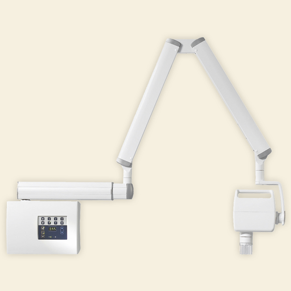
Miniray
Intra-oral radiological unitis a reliable system thanks to superior image quality and the validity of diagnostic results.
The high frequency and constant potential generator enhance the image quality and are not affected by voltage variations in the power supply line. Compared to the AC generators, the constant potential technology allows to generate more energized and harder radiations, reducing the soft ones.
MINIRAY is compatible with all digital radiovideography systems based on IP sensors technology (e.g. DIGORA) or CCD.
MINIRAY is equipped with an articulated arm that allows the head of the device to be kept in position during exposure. The practical handle of the exposure head allows its quick and easy single-handed maneuvering.
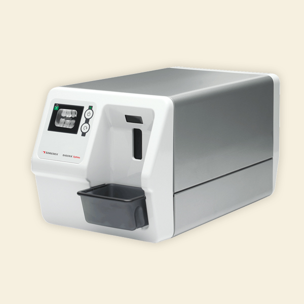
Digora
DIGORA OPTIME UV represents the latest evolution in the field of digital phosphor imaging. With the UV model, SOREDEX introduces further improvements in the already excellent performance of the Digora Optime system.
In this new model, the technical characteristics inherent to image quality are further improved: 14.3 pairs of lines per millimeter, combined with the high dynamic range, guarantee precise, sharp images of high diagnostic value.
The new DIGORA OPTIME UV system introduces new automatic image optimization functions, capable of guaranteeing radiographs perfectly calibrated on the gray scale.
This innovative system, patented by Soredex, allows 99.9% decontamination of digital phosphor plates during the reading phase. Viruses and bacteria such as HIV, hepatitis, herpes and many others are eradicated by the new OPTICLEAN system, guaranteeing high levels of safety for both patients and, above all, the staff, who ismore frequently exposed to cross-contamination. The system, through a particular technique, patented and certified by accredited university bodies, uses ultraviolet light to decontaminate the entire surface of the plate, returning it safe and perfectly decontaminated after reading.
In the DIGORA OPTIME UV system,the plates are secured with plate covers and safety protections to ensure optimal control and prevention of cross-contamination between platelets, the reader and external protections.
Contact us to book an examination immediately
Book examination